

Search results
Search for "sugars" in Full Text gives 196 result(s) in Beilstein Journal of Organic Chemistry.
Recent advances in the gold-catalyzed additions to C–C multiple bonds
- He Huang,
- Yu Zhou and
- Hong Liu
Beilstein J. Org. Chem. 2011, 7, 897–936, doi:10.3762/bjoc.7.103
- tetrasaccharide 74, which demonstrated its versatility and efficacy. Another study [43] showed that 1,6-anhydro sugars 76 and 78 could be synthesized by utilizing salient features of gold-catalyzed glycosidations. 2.2 Aldehydes and ketones as nucleophiles Different oxygen heterocycles can be obtained from the

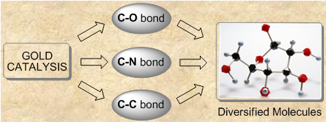
Graphical Abstract

Scheme 1: Gold-catalyzed addition of alcohols.
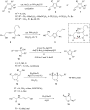
Scheme 2: Gold-catalyzed cycloaddition of alcohols.

Scheme 3: Ionic liquids as the solvent in gold-catalyzed cycloaddition.

Scheme 4: Gold-catalyzed cycloaddition of diynes.

Scheme 5: Gold(I) chloride catalyzed cycloisomerization of 2-alkynyl-1,5-diols.

Scheme 6: Gold-catalyzed cycloaddition of glycols and dihydroxy compounds.

Scheme 7: Gold-catalyzed ring-opening of cyclopropenes.

Scheme 8: Gold-catalyzed intermolecular hydroalkoxylation of alkynes. PR3 = 41–45.

Scheme 9: Gold-catalyzed intramolecular 6-endo-dig cyclization of β-hydroxy-α,α-difluoroynones.

Scheme 10: Gold-catalyzed intermolecular hydroalkoxylation of non-activated olefins.

Scheme 11: Preparation of unsymmetrical ethers from alcohols.

Scheme 12: Expedient synthesis of dihydrofuran-3-ones.

Scheme 13: Catalytic approach to functionalized divinyl ketones.
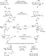
Scheme 14: Gold-catalyzed glycosylation.

Scheme 15: Gold-catalyzed cycloaddition of aldehydes and ketones.

Scheme 16: Gold-catalyzed annulations of 2-(ynol)aryl aldehydes and o-alkynyl benzaldehydes.

Scheme 17: Gold-catalyzed addition of carboxylates.

Scheme 18: Dual-catalyzed rearrangement reaction of allenoates.

Scheme 19: Meyer–Schuster rearrangement of propargylic alcohols.

Scheme 20: Propargylic alcohol rearrangements.

Scheme 21: Gold-catalyzed synthesis of imines and amine alkylation.

Scheme 22: Hydroamination of allenes and allenamides.

Scheme 23: Gold-catalyzed inter- and intramolecular amination of alkynes and alkenes.

Scheme 24: Gold-catalyzed cycloisomerization of O-propioloyl oximes and β-allenylhydrazones.

Scheme 25: Intra- and intermolecular amination with ureas.

Scheme 26: Gold-catalyzed cyclization of ortho-alkynyl-N-sulfonylanilines and but-3-yn-1-amines.

Scheme 27: Gold-catalyzed piperidine ring synthesis.

Scheme 28: Ring expansion of alkylnyl cyclopropanes.

Scheme 29: Gold-catalyzed annulations of N-propargyl-β-enaminones and azomethine imines.
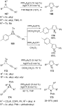
Scheme 30: Gold(I)-catalyzed cycloisomerization of aziridines.

Scheme 31: AuCl3/AgSbF6-catalyzed intramolecular amination of 2-(tosylamino)phenylprop-1-en-3-ols.

Scheme 32: Gold-catalyzed cyclization via a 7-endo-dig pathway.

Scheme 33: Gold-catalyzed synthesis of fused xanthines.

Scheme 34: Gold-catalyzed synthesis of amides and isoquinolines.

Scheme 35: Gold-catalyzed oxidative cross-coupling reactions of propargylic acetates.

Scheme 36: Gold-catalyzed nucleophilic addition to allenamides.

Scheme 37: Gold-catalyzed direct carbon–carbon bond coupling reactions.

Scheme 38: Gold-catalyzed C−H functionalization of indole/pyrrole heterocycles and non-activated arenes.

Scheme 39: Gold-catalyzed cycloisomerization of cyclic compounds.

Scheme 40: Gold-catalyzed cycloaddition of 1-aryl-1-allen-6-enes and propargyl acetates.

Scheme 41: Gold(I)-catalyzed cycloaddition with ligand-controlled regiochemistry.

Scheme 42: Gold(I)-catalyzed cycloaddition of dienes and enynes.

Scheme 43: Gold-catalyzed intramolecular cycloaddition of 3-alkoxy-1,5-enynes and 2,2-dipropargylmalonates.

Scheme 44: Gold-catalyzed intramolecular cycloaddition of 1,5-allenynes.

Scheme 45: Gold(I)-catalyzed cycloaddition of indoles.

Scheme 46: Gold-catalyzed annulation reactions.

Scheme 47: Gold–carbenoid induced cleavage of a sp3-hybridized C−H bond.

Scheme 48: Furan- and indole-based cascade reactions.

Scheme 49: Tandem process using aromatic alkynes.

Scheme 50: Gold-catalyzed cycloaddition of 1,3-dien-5-ynes.

Scheme 51: Gold-catalyzed cascade cyclization of diynes, propargylic esters, and 1,3-enynyl ketones.

Scheme 52: Tandem reaction of β-phenoxyimino ketones and alkynyl oxime ethers.

Scheme 53: Gold-catalyzed tandem cyclization of enynes, 2-(tosylamino)phenylprop-1-yn-3-ols, and allenoates.

Scheme 54: Cyclization of 2,4-dien-6-yne carboxylic acids.
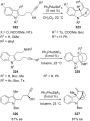
Scheme 55: Gold(I)-catalyzed tandem cyclization approach to tetracyclic indolines.
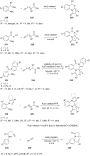
Scheme 56: Gold-catalyzed tandem reactions of alkynes.
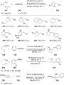
Scheme 57: Aminoarylation and oxyarylation of alkenes.

Scheme 58: Cycloaddition of 2-ethynylnitrobenzene with various alkenes.

Scheme 59: Gold-catalyzed tandem reactions of allenoates and alkynes.

Scheme 60: Gold-catalyzed asymmetric synthesis of 2,3-dihydropyrroles.

Scheme 61: Chiral [NHC–Au(I)]-catalyzed cyclization of enyne.
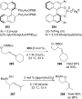
Scheme 62: Gold-catalyzed hydroaminations and hydroalkoxylations.

Scheme 63: Gold(I)-catalyzed asymmetric hydroalkoxylation of 1,3-dihydroxymethyl-2-alkynylbenzene chromium com...

Scheme 64: Gold-catalyzed synthesis of julolidine derivatives.

Scheme 65: Gold-catalyzed the synthesis of chiral fused heterocycles.

Scheme 66: Gold-catalyzed asymmetric reactions with 3,5-(t-Bu)2-4-MeO-MeOBIPHEP.

Scheme 67: Gold-catalyzed cyclization of o-(alkynyl) styrenes.

Scheme 68: Asymmetric gold(I)-catalyzed redox-neutral domino reactions of enynes.

Scheme 69: Gold(I)-catalyzed enantioselective polyene cyclization reaction.

Scheme 70: Gold(I)-catalyzed enantioselective synthesis of benzopyrans.

Scheme 71: Gold(I)-catalyzed enantioselective ring expansion of allenylcyclopropanols.
Metathesis access to monocyclic iminocyclitol-based therapeutic agents
- Ileana Dragutan,
- Valerian Dragutan,
- Carmen Mitan,
- Hermanus C.M. Vosloo,
- Lionel Delaude and
- Albert Demonceau
Beilstein J. Org. Chem. 2011, 7, 699–716, doi:10.3762/bjoc.7.81

- , can be considered as carbohydrate mimics in which the endocyclic oxygen atom of sugars has been replaced by an imino group. This vast and highly diversified class has attracted considerable interest due to the remarkable biological profile shown by many of its members which has been detailed in a
- number of excellent books and reviews [1][2][3][4][5][6][7][8][9][10][11][12]. Natural iminosugars (i.e., alkaloids mimicking the structures of sugars, widespread in many plants or microorganisms) [12][13][14][15], as well as non-natural counterparts, are becoming important leads for drug development in

Graphical Abstract

Scheme 1: Well-defined Mo- and Ru-alkylidene metathesis catalysts.

Scheme 2: Representative pyrrolidine-based iminocyclitols.

Scheme 3: Synthesis of (±)-(2R*,3R*,4S*)-2-hydroxymethylpyrrolidin-3,4-diol (18), (±)-2-hydroxymethylpyrrolid...

Scheme 4: Synthesis of enantiopure iminocyclitol (−)-(2S,3R,4S,5S)-2,5-dihydroxymethylpyrrolidin-3,4-diol (23...

Scheme 5: Synthesis of 1,4-dideoxy-1,4-imino-D-allitol (29) and formal synthesis of (2S,3R,4S)-3,4-dihydroxyp...

Scheme 6: Synthesis of iminocyclitols 35 and 36.

Scheme 7: Total synthesis of iminocyclitols 40 and 44.

Scheme 8: Synthesis of 2,5-dideoxy-2,5-imino-D-mannitol [(+)-DMDP] (49) and (−)-bulgecinine (50).

Scheme 9: Synthesis of (+)-broussonetine G (53).
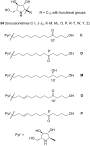
Scheme 10: Structural features of broussonetines 54.

Scheme 11: Synthesis of broussonetines by cross-metathesis.
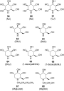
Scheme 12: Representative piperidine-based iminocyclitols.

Scheme 13: Total synthesis of 1-deoxynojirimycin (62) and 1-deoxyaltronojirimycin (65).

Scheme 14: Synthesis by RCM of 1-deoxymannonojirimycin (63) and 1-deoxyallonojirimycin (66).

Scheme 15: Total synthesis of (+)-1-deoxynojirimycin (62).

Scheme 16: Synthesis of ent-1,6-dideoxynojirimycin (83) and 5-amino-1,5,6-trideoxyaltrose (84).

Scheme 17: Synthesis of 1-deoxygalactonojirimycin (64), 1-deoxygulonojirimycin (91) and 1-deoxyidonojirimycin (...

Scheme 18: Synthesis of L-1-deoxyaltronojirimycin (96).

Scheme 19: Synthesis of 1-deoxymannonojirimycin (63) and 1-deoxyaltronojirimycin (65).

Scheme 20: Synthesis of 5-des(hydroxymethyl)-1-deoxymannonojirimycin (111) and 5-des(hydroxymethyl)-1-deoxynoj...

Scheme 21: Synthesis of D-1-deoxygulonojirimycin (91) and L-1-deoxyallonojirimycin (122).

Scheme 22: Total synthesis of fagomine (129), 3-epi-fagomine (126) and 3,4-di-epi-fagomine (130).

Scheme 23: Total synthesis of (+)-adenophorine (135).

Scheme 24: Total synthesis of (+)-5-deoxyadenophorine (138) and analogues 142–145.

Scheme 25: Synthesis by RCM of 1,6-dideoxy-1,6-iminoheptitols 148 and 149.

Scheme 26: Synthesis by RCM of oxazolidinyl azacycles 152 and 154.

Scheme 27: Representative azepane-based iminocyclitols.

Scheme 28: Synthesis of hydroxymethyl-1-(4-methylphenylsulfonyl)azepane 3,4,5-triol (169).

Scheme 29: Synthesis by RCM of tetrahydropyridin-3-ol 171 and tetrahydroazepin-3-ol 173.
Synthesis of glycoconjugate fragments of mycobacterial phosphatidylinositol mannosides and lipomannan
- Benjamin Cao,
- Jonathan M. White and
- Spencer J. Williams
Beilstein J. Org. Chem. 2011, 7, 369–377, doi:10.3762/bjoc.7.47
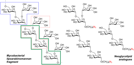
- of a 3,4-butanediacetal-protected mannoside. The azidooctyl groups of these synthetic mannans were elaborated to fluorescent glycoconjugates and squaric ester derivatives useful for further conjugation studies. Keywords: fluorescently-labelled sugars; glycoconjugates; lipomannan; mycobacteria

Graphical Abstract

Scheme 1: Indicative topology model for the biosynthesis of the glycophospholipids PIMs, LM and LAM in mycoba...

Figure 1: Chemical structures of (A) a representative PIM, AcPIM5 and (B) a mannan fragment of LM from mycoba...

Figure 2: Target di- and trisaccharide glycoconjugate fragments of PIMs and LM.

Scheme 2: Synthesis of azidooctyl alcohol 14 and diol 15.

Scheme 3: Synthesis of mannosyl donors 16 and 17.

Figure 3: ORTEP plot of single crystal X-ray determination of (7S)-18. Thermal ellipsoids denote 20% electron...

Scheme 4: Synthesis of disaccharide 1 and trisaccharide 2.

Scheme 5: Synthesis of trisaccharide 3.

Scheme 6: Synthesis of trisaccharide 4.

Scheme 7: Synthesis of glycoconjugates 5–7 and 8–10.
Preparation of aminoethyl glycosides for glycoconjugation
- Robert Šardzík,
- Gavin T. Noble,
- Martin J. Weissenborn,
- Andrew Martin,
- Simon J. Webb and
- Sabine L. Flitsch
Beilstein J. Org. Chem. 2010, 6, 699–703, doi:10.3762/bjoc.6.81

- general aminoethylation method that was applicable to all targets was found, which rather suggests that protocols need to be tailored for each sugar. Results and Discussion Coupling reactions Aminoethyl glycosides have previously been generated in a number of ways. Free sugars have been glycosylated with
- ], bromoethanol [16] or azidoethanol [17] and subsequently transformed into the amine. In the interest of finding fast reliable methods, we have investigated two general aminoethylation protocols: First, the direct glycosylation of peracetylated sugars, which can be either purchased or easily prepared from free
- sugars and can be used without purification. Where these proved to be unreactive, the anomeric acetates were converted to glycosyl bromides, usually in quantitative yields, and products were used without further purification. Where possible, N-Cbz-protected aminoethanol was used as the glycosyl acceptor

Graphical Abstract

Figure 1: Aminoethyl glycosides (1–9) which were synthesised in this study.

Scheme 1: General reaction scheme for generation of aminoethyl glycosides. X = OAc, Br or Cl.

Scheme 2: Deprotection protocols.
Addition of lithiated enol ethers to nitrones and subsequent Lewis acid induced cyclizations to enantiopure 3,6-dihydro-2H-pyrans – an approach to carbohydrate mimetics
- Fabian Pfrengle and
- Hans-Ulrich Reissig
Beilstein J. Org. Chem. 2010, 6, No. 75, doi:10.3762/bjoc.6.75
- ]. A final hydrogenation step gave aminopyran 30 in high overall yield. Aminopyrans 24, 26, 28 and 30 can be regarded as mimetics of differently configured (2-deoxy) 4-amino sugars [24][25][26]. Aminopyrans 24 and 28 have already been linked via amide bonds to the thiol shell of gold nanoparticles

Graphical Abstract

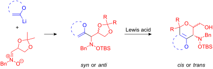
Scheme 1: Synthesis of 3,6-dihydro-2H-pyrans D.

Scheme 2: Addition of lithiated enol ether 1a to nitrone 2c/Et2AlCl.

Scheme 3: Mechanistic proposal for the transformation of syn-4a into cis-5a in the presence of TMSOTf.

Scheme 4: Unsuccessful attempt to cyclize hydroxylamine derivative syn-4f.

Scheme 5: Functionalizations of the enol ether moiety of cis-5a leading to compounds 8–11.

Scheme 6: Transformations of the enol ether moiety of trans-5a leading to products 13–15.

Scheme 7: Bromination of bicyclic dihydropyran trans-5b affording 16.

Scheme 8: Synthesis of epoxypyran 18 by bromination of cis-5d.

Scheme 9: Oxidative cleavage of dihydropyran 19 to lactone 20.

Scheme 10: Transformation of α-bromoketone 15 into nitrone 21 by an internal redox reaction.

Scheme 11: Transformation of α-bromoketone 11 into nitrone 21.

Scheme 12: Synthesis of diastereomeric aminopyrans 24 and 26.

Scheme 13: Synthesis of diastereomeric aminopyrans 28 and 30.

Scheme 14: Synthesis of triamides 31 and 32.
New amphiphilic glycopolymers by click functionalization of random copolymers – application to the colloidal stabilisation of polymer nanoparticles and their interaction with concanavalin A lectin
- Otman Otman,
- Paul Boullanger,
- Eric Drockenmuller and
- Thierry Hamaide
Beilstein J. Org. Chem. 2010, 6, No. 58, doi:10.3762/bjoc.6.58
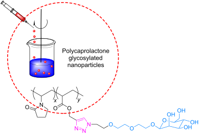
- drawbacks inherent to polymer structures. Due to the high versatility of click chemistry in the presence of functional groups, it was then of interest to attempt the reaction with unprotected sugars. Moreover, this chemistry can be carried out not only in various organic solvents, but also in aqueous

Graphical Abstract

Figure 1: Preparation of the 8-azido-3,6-dioxaoctyl α-D-mannopyranoside.

Figure 2: Preparation of poly(propargyl-co-N-vinyl pyrrolidone) and subsequent addition of the mannose deriva...

Figure 3: Size of the nanoparticles stabilized with Pluronic® F-68/NVP-PA-Man (0.8/0.2), after addition of in...

Figure 4: Hydrogen and carbon numbering for NMR assignment.
Synthesis of glycosylated β3-homo-threonine conjugates for mucin-like glycopeptide antigen analogues
- Florian Karch and
- Anja Hoffmann-Röder
Beilstein J. Org. Chem. 2010, 6, No. 47, doi:10.3762/bjoc.6.47
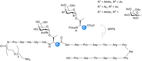
- instance, stable TACA mimics comprising C-glycosides [11][12][13][14], S-glycosides [15][16][17][18][19] and deoxyfluoro sugars [20] have been used to circumvent hydrolytic degradation by endogenous glycosidases. In principle, antigenicity of the artificial TACA derivatives should be enhanced by minor

Graphical Abstract

Figure 1: Structures of the naturally occurring TN and TF antigens and the targeted Fmoc-β3hThr analogues.

Scheme 1: Synthesis of Fmoc-β3hThr antigen conjugates by Arndt–Eistert homologation.

Scheme 2: Solid-phase synthesis of the tumour-associated MUC1 α/β-hybrid glycopeptide analogue 8 carrying the...
Molecular recognition of organic ammonium ions in solution using synthetic receptors
- Andreas Späth and
- Burkhard König
Beilstein J. Org. Chem. 2010, 6, No. 32, doi:10.3762/bjoc.6.32

Graphical Abstract

Figure 1: Biologically important amines and quaternary ammonium salts: histamine (1), dopamine (2) and acetyl...

Figure 2: Crown ether 18-crown-6.

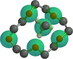
Figure 3: Conformations of 18-crown-6 (4) in solvents of different polarity.

Figure 4: Binding topologies of the ammonium ion depending on the crown ring size.
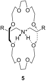
Figure 5: A “pseudorotaxane” structure consisting of 24-crown-8 and a secondary ammonium ion (5); R = Ph.

Figure 6: Typical examples of azacrown ethers, cryptands and related aza macrocycles.

Figure 7: Binding of ammonium to azacrown ethers and cryptands [111-113].

Figure 8: A 19-crown-6-ether with decalino blocking groups (11) and a thiazole-dibenzo-18-crown-6-ether (12).

Figure 9: 1,3-Bis(6-oxopyridazin-1-yl)propane derivatives 13 and 14 by Campayo et al.

Figure 10: Fluorescent azacrown-PET-sensors based on coumarin.

Figure 11: Two different pyridino-cryptands (17 and 18) compared to a pyridino-crown (19); chiral ammonium ion...

Figure 12: Pyridino-18-crown-6 ligand (21), a similar acridino-18-crown-6 ligand (22) and a structurally relat...

Figure 13: Ciral pyridine-azacrown ether receptors 24.

Figure 14: Chiral 15-crown-5 receptors 26 and an analogue 18-crown-6 ligand 27 derived from amino alcohols.

Figure 15: C2-symmetric chiral 18-crown-6 amino alcohol derivatives 28 and related macrocycles.

Figure 16: Macrocycles with diamide-diester groups (30).

Figure 17: C2-symmetric chiral aza-18-crown-6 ethers (31) with phenethylamine residues.

Figure 18: Chiral C-pivot p-methoxy-phenoxy-lariat ethers.

Figure 19: Chiral lariat crown ether 34.

Figure 20: Sucrose-based chiral crown ether receptors 36.
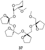
Figure 21: Permethylated fructooligosaccharide 37 showing induced-fit chiral recognition.

Figure 22: Biphenanthryl-18-crown-6 derivative 38.

Figure 23: Chiral lariat crown ethers derived from binol by Fuji et al.

Figure 24: Chiral phenolic crown ether 41 with “aryl chiral barriers” and guest amines.
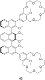
Figure 25: Chiral bis-crown receptor 43 with a meso-ternaphthalene backbone.

Figure 26: Chromogenic pH-dependent bis-crown chemosensor 44 for diamines.

Figure 27: Triamine guests for binding to receptor 44.

Figure 28: Chiral bis-crown phenolphthalein chemosensors 46.

Figure 29: Crown ether amino acid 47.

Figure 30: Luminescent receptor 48 for bis-alkylammonium guests.

Figure 31: Luminescent CEAA (49a), a bis-CEAA receptor for amino acids (49b) and the structure of lysine bindi...

Figure 32: Luminescent CEAA tripeptide for binding small peptides.

Figure 33: Bis crown ether 51a self assembles co-operatively with C60-ammonium ion 51b.

Figure 34: Triptycene-based macrotricyclic dibenzo-[24]-crown-8 ether host 52 and guests.

Figure 35: Copper imido diacetic acid azacrown receptor 53a and the suggested His-Lys binding motif; a copper ...

Figure 36: Urea (54) and thiourea (55) benzo crown receptor for transport and extraction of amino acids.

Figure 37: Crown pyryliums ion receptors 56 for amino acids.

Figure 38: Ditopic sulfonamide bridged crown ether receptor 57.

Figure 39: Luminescent peptide receptor 58.

Figure 40: Luminescent receptor 59 for the detection of D-glucosamine hydrochloride in water/ethanol and lumin...

Figure 41: Guanidinium azacrown receptor 61 for simple amino acids and ditopic receptor 62 with crown ether an...

Figure 42: Chiral bicyclic guanidinium azacrown receptor 63 and similar receptor 64 for the enantioselective t...

Figure 43: Receptors for zwitterionic species based on luminescent CEAAs.

Figure 44: 1,10-Azacrown ethers with sugar podand arms and the anticancer agent busulfan.

Figure 45: Benzo-18-crown-6 modified β-cyclodextrin 69 and β-cyclodextrin functionalized with diaza-18-crown-6...

Figure 46: Receptors for colorimetric detection of primary and secondary ammonium ions.

Figure 47: Porphyrine-crown-receptors 72.

Figure 48: Porphyrin-crown ether conjugate 73 and fullerene-ammonium ion guest 74.

Figure 49: Calix[4]arene (75a), homooxocalix[4]arene (75b) and resorcin[4]arene (75c) compared (R = H, alkyl c...

Figure 50: Calix[4]arene and ammonium ion guest (R = H, alkyl, OAcyl etc.), possible binding sites; A: co-ordi...

Figure 51: Typical guests for studies with calixarenes and related molecules.
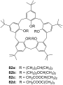
Figure 52: Lower rim modified p-tert-butylcalix[5]arenes 82.

Figure 53: The first example of a water soluble calixarene.

Figure 54: Sulfonated water soluble calix[n]arenes that bind ammonium ions.
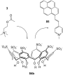
Figure 55: Displacement assay for acetylcholine (3) with a sulfonato-calix[6]arene (84b).

Figure 56: Amino acid inclusion in p-sulfonatocalix[4]arene (84a).

Figure 57: Calixarene receptor family 86 with upper and lower rim functionalization.

Figure 58: Calix[6]arenes 87 with one carboxylic acid functionality.
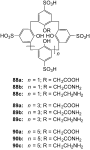
Figure 59: Sulfonated calix[n]arenes with mono-substitution at the lower rim systematically studied on their r...

Figure 60: Cyclotetrachromotropylene host (91) and its binding to lysine (81c).

Figure 61: Calixarenes 92 and 93 with phosphonic acids groups.

Figure 62: Calix[4]arene tetraphosphonic acid (94a) and a double bridged analogue (94b).
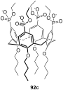
Figure 63: Calix[4]arene tetraphosphonic acid ester (92c) for surface recognition experiments.

Figure 64: Calixarene receptors 95 with α-aminophosphonate groups.

Figure 65: A bridged homocalix[3]arene 95 and a distally bridged homocalix[4]crown 96.

Figure 66: Homocalix[3]arene ammonium ion receptor 97a and the Reichardt’s dye (97b) for colorimetric assays.

Figure 67: Chromogenic diazo-bridged calix[4]arene 98.

Figure 68: Calixarene receptor 99 by Huang et al.
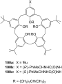
Figure 69: Calixarenes 100 reported by Parisi et al.

Figure 70: Guest molecules for inclusion in calixarenes 100: DAP × 2 HCl (101a), APA (101b) and Lys-OMe × 2 HC...

Figure 71: Different N-linked peptido-calixarenes open and with glycol chain bridges.

Figure 72: (S)-1,1′-Bi-2-naphthol calixarene derivative 104 published by Kubo et al.

Figure 73: A chiral ammonium-ion receptor 105 based on the calix[4]arene skeleton.

Figure 74: R-/S-phenylalaninol functionalized calix[6]arenes 106a and 106b.

Figure 75: Capped homocalix[3]arene ammonium ion receptor 107.

Figure 76: Two C3 symmetric capped calix[6]arenes 108 and 109.

Figure 77: Phosphorous-containing rigidified calix[6]arene 110.

Figure 78: Calix[6]azacryptand 111.

Figure 79: Further substituted calix[6]azacryptands 112.

Figure 80: Resorcin[4]arene (75c) and the cavitands (113).

Figure 81: Tetrasulfonatomethylcalix[4]resorcinarene (114).
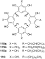
Figure 82: Resorcin[4]arenes (115a/b) and pyrogallo[4]arenes (115c, 116).
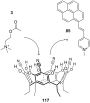
Figure 83: Displacement assay for acetylcholine (3) with tetracyanoresorcin[4]arene (117).

Figure 84: Tetramethoxy resorcinarene mono-crown-5 (118).

Figure 85: Components of a resorcinarene based displacement assay for ammonium ions.

Figure 86: Chiral basket resorcin[4]arenas 121.

Figure 87: Resorcinarenes with deeper cavitand structure (122).

Figure 88: Resorcinarene with partially open deeper cavitand structure (123).
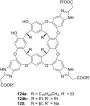
Figure 89: Water-stabilized deep cavitands with partially structure (124, 125).
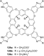
Figure 90: Charged cavitands 126 for tetralkylammonium ions.

Figure 91: Ditopic calix[4]arene receptor 127 capped with glycol chains.

Figure 92: A calix[5]arene dimer for diammonium salt recognition.

Figure 93: Calixarene parts 92c and 129 for the formation molecular capsules.
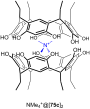
Figure 94: Encapsulation of a quaternary ammonium cation by two resorcin[4]arene molecules (NMe4+@[75c]2 × Cl−...

Figure 95: Encapsulation of a quaternary ammonium cation by six resorcin[4]arene molecules (NMe3D+@[130]6 × Cl−...

Figure 96: Structure and schematic of cucurbit[6]uril (CB[6], 131a).
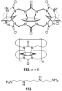
Figure 97: Cyclohexanocucurbit[6]uril (CB′[6], 132) and the guest molecule spermine (133).

Figure 98: α,α,δ,δ-Tetramethylcucurbit[6]uril (134).

Figure 99: Structure of the cucurbituril-phthalhydrazide analogue 135.

Figure 100: Organic cavities for the displacement assay for amine differentiation.

Figure 101: Displacement assay methodology for diammonium- and related guests involving cucurbiturils and some ...

Figure 102: Nor-seco-Cucurbituril (±)-bis-ns-CB[6] (140) and guest molecules.

Figure 103: The cucurbit[6]uril based complexes 141 for chiral discrimination.
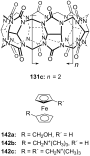
Figure 104: Cucurbit[7]uril (131c) and its ferrocene guests (142) opposed.

Figure 105: Cucurbit[7]uril (131c) guest inclusion and representative guests.

Figure 106: Cucurbit[7]uril (131c) binding to succinylcholine (145) and different bis-ammonium and bis-phosphon...

Figure 107: Paraquat-cucurbit[8]uril complex 149.
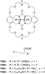
Figure 108: Gluconuril-based ammonium receptors 150.
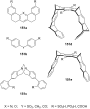
Figure 109: Examples of clefts (151a), tweezers (151b, 151c, 151d) and clips (151e).

Figure 110: Kemp’s triacid (152a), on example of Rebek’s receptors (152b) and guests.

Figure 111: Amino acid receptor (154) by Rebek et al.

Figure 112: Hexagonal lattice designed hosts by Bell et al.

Figure 113: Bell’s amidinium receptor (156) and the amidinium ion (157).
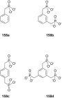
Figure 114: Aromatic phosphonic acids.

Figure 115: Xylene phosphonates 159 and 160a/b for recognition of amines and amino alcohols.
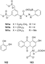
Figure 116: Bisphosphonate recognition motif 161 for a colorimetric assay with alizarin complexone (163) for ca...

Figure 117: Bisphosphonate/phosphate clip 164 and bisphosphonate cleft 165.

Figure 118: N-Methylpyrazine 166a, N-methylnicotinamide iodide (166b) and NAD+ (166c).

Figure 119: Bisphosphate cavitands.
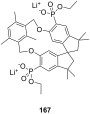
Figure 120: Bisphosphonate 167 of Schrader and Finocchiaro.
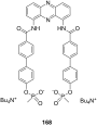
Figure 121: Tweezer 168 for noradrenaline (80b).

Figure 122: Different tripods and heparin (170).
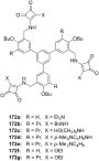
Figure 123: Squaramide based receptors 172.
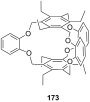
Figure 124: Cage like NH4+ receptor 173 of Kim et al.

Figure 125: Ammonium receptors 174 of Chin et al.

Figure 126: 2-Oxazolin-based ammonium receptors 175a–d and 176 by Ahn et al.

Figure 127: Racemic guest molecules 177.

Figure 128: Tripods based on a imidazole containing macrocycle (178) and the guest molecules employed in the st...

Figure 129: Ammonium ion receptor 180.

Figure 130: Tetraoxa[3.3.3.3]paracyclophanes 181 and a cyclophanic tetraester (182).

Figure 131: Peptidic bridged paraquat-cyclophane.
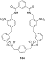
Figure 132: Shape-selective noradrenaline host.

Figure 133: Receptor 185 for binding of noradrenaline on surface layers from Schrader et al.

Figure 134: Tetraphosphonate receptor for binding of noradrenaline.

Figure 135: Tetraphosphonate 187 of Schrader and Finocchiaro.

Figure 136: Zinc-Porphyrin ammonium-ion receptors 188 and 189 of Mizutani et al.
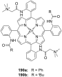
Figure 137: Zinc porphyrin receptor 190.

Figure 138: Zinc porphyrin receptors 191 capable of amino acid binding.

Figure 139: Zinc-porphyrins with amino acid side chains for stereoinduction.

Figure 140: Bis-zinc-bis-porphyrin based on Tröger’s base 193.

Figure 141: BINAP-zinc-prophyrin derivative 194 and it’s guests.

Figure 142: Bisaryl-linked-zinc-porphyrin receptors.
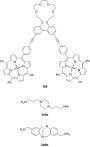
Figure 143: Bis-zinc-porphyrin 199 for diamine recognition and guests.

Figure 144: Bis-zinc-porphyrin crown ether 201.

Figure 145: Bis-zinc-porphyrin 202 for stereodiscrimination (L = large substituent; S = small substituent).

Figure 146: Bis-zinc-porphyrin[3]rotaxane and its copper complex and guests.

Figure 147: Dien-bipyridyl ligand 206 for co-ordination of two metal atoms.

Figure 148: The ligand and corresponding tetradentate co-complex 207 serving as enantioselective receptor for a...

Figure 149: Bis(oxazoline)–copper(II) complex 208 for the recognition of amino acids in aqueous solution.

Figure 150: Zinc-salen-complexes 209 for the recognition tertiary amines.

Figure 151: Bis(oxazoline)–copper(II) 211 for the recognition of amino acids in aqueous solution.

Figure 152: Zn(II)-complex of a C2 terpyridine crown ether.

Figure 153: Displacement assay and receptor for aspartate over glutamate.

Figure 154: Chiral complex 214 for a colorimetric displacement assay for amino acids.

Figure 155: Metal complex receptor 215 with tripeptide side arms.

Figure 156: A sandwich complex 216 and its displaceable dye 217.

Figure 157: Lanthanide complexes 218–220 for amino acid recognition.
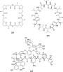
Figure 158: Nonactin (221), valinomycin (222) and vancomycin (223).

Figure 159: Monesin (224a) and a chiral analogue for enantiodiscrimination of ammonium guests (224b).

Figure 160: Chiral podands (226) compared to pentaglyme-dimethylether (225) and 18-crown-6 (4).

Figure 161: Lasalocid A (228).

Figure 162: Lasalocid derivatives (230) of Sessler et al.

Figure 163: The Coporphyrin I tetraanion (231).

Figure 164: Linear and cyclic peptides for ammonium ion recognition.

Figure 165: Cyclic and bicyclic depsipeptides for ammonium ion recognition.
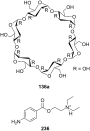
Figure 166: α-Cyclodextrin (136a) and novocaine (236).
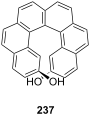
Figure 167: Helical diol receptor 237 by Reetz and Sostmann.

Figure 168: Ammonium binding spherand by Cram et al. (238a) and the cyclic[6]metaphenylacetylene 238b in compar...

Figure 169: Receptor for peptide backbone and ammonium binding (239).

Figure 170: Anion sensor principle with 3-hydroxy-2-naphthanilide of Jiang et al.

Figure 171: 7-bromo-3-hydroxy-N-(2-hydroxyphenyl)naphthalene 2-carboxamide (241) and its amine binding.

Figure 172: Naturally occurring catechins with affinity to quaternary ammonium ions.

Figure 173: Spiropyran (244) and merocyanine form (244a) of the amino acid receptors of Fuji et al.

Figure 174: Coumarin aldehyde (245) and its iminium species with amino acid bound (245a) by Glass et al.
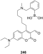
Figure 175: Coumarin aldehyde appended with boronic acid.

Figure 176: Quinolone aldehyde dimers by Glass et al.

Figure 177: Chromogenic ammonium ion receptors with trifluoroacetophenone recognition motifs.

Figure 178: Chromogenic ammonium ion receptor with trifluoroacetophenone recognition motif bound on different m...
Bioorthogonal metabolic glycoengineering of human larynx carcinoma (HEp-2) cells targeting sialic acid
- Arne Homann,
- Riaz-ul Qamar,
- Sevnur Serim,
- Petra Dersch and
- Jürgen Seibel
Beilstein J. Org. Chem. 2010, 6, No. 24, doi:10.3762/bjoc.6.24
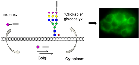
- any significant background fluorescence (Figure 1). In both the Neu5Hex fed HEp-2 and the incubation with Ac4GlcNAz a clear staining of the cellular glycocalyx at the expected wavelengths was observed. Conclusion Sialic acids are prominent sugars which are located in the terminal position on cell

Graphical Abstract

Scheme 1: The natural forms of sialic acids, human N-acetylneuraminic acid (Neu5Ac, 1) and mammalian N-glycol...

Scheme 2: Synthesis of N-(1-oxohex-5-ynyl)neuraminic acid (Neu5Hex 3).
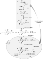
Scheme 3: Metabolic pathway of Ac4GlcNAz and the genetic control of Neu5Ac 1 synthesis by feedback inhibition...

Scheme 4: Proposed metabolic pathway of Neu5Hex 3 based on known mechanisms of Neu5Gc 2 uptake [5]. TGN: trans-G...

Scheme 5: Labelling of alkynylated neuraminic acid by azido-fluorescein.

Figure 1: Top left: HEp-2 cells incorporated with Ac4GlcNAz 16, labelled with alkynylated TAMRA at 580 nm. Bo...
(Pseudo)amide-linked oligosaccharide mimetics: molecular recognition and supramolecular properties
- José L. Jiménez Blanco,
- Fernando Ortega-Caballero,
- Carmen Ortiz Mellet and
- José M. García Fernández
Beilstein J. Org. Chem. 2010, 6, No. 20, doi:10.3762/bjoc.6.20
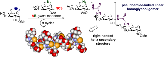
- ; glycomimetics; pseudooligosaccharides; spaced sugars; Review Among the major classes of biomolecules, carbohydrates are characterized by nearly unlimited structural diversity. Monosaccharide units can combine to produce oligosaccharides in a number of permutations that increases rapidly with the number of
- ]. In addition, SAAs have been widely used in the field of material sciences as carbohydrate-derived monomers for the design of novel chiral polyamides [18]. Nevertheless, these review articles mainly focus on amide-linked sugars and do not pay too much attention to other types of pseudosugars. A
- notable exception is the contribution by Wessel and Dias Lucas, who recently published an interesting review where oligosaccharide mimetics which deviate from the natural linking pattern were discussed [19]. This review also included pseudoamide-linked sugars (Figure 1). In this current review we have

Graphical Abstract

Figure 1: Schematic representation of sugar aminoacids (SAAs) and (pseudo)amide oligosaccharide mimetics.

Figure 2: Natural SAAs structures and natural nucleosidic antibiotics.

Scheme 1: Synthetic route to the target amide-linked sialooligomers. (a) Fmoc-Cl, NaHCO3, H2O, dioxane, 0 °C....

Figure 3: The general structure of glycoamino acids and their corresponding oligomers.

Figure 4: Conformational analysis of the β(1→2)-amide-linked glucooligomer 9.

Figure 5: Short oligomeric chains of C-glycosyl D-arabino THF amino acid oligomers.

Figure 6: (A) Stereoview of the minimized structure of compound 16 (produced by a 500 ps simulation) that mos...

Figure 7: Structures of linear oxetane-β- and δ-SAA homo-oligomers 19–20.

Figure 8: 10-Membered ring H-bonds in compound 21 consistent with NMR and modelling investigations.

Figure 9: General structure of carbopeptoid-oligonucleotide conjugates.

Figure 10: Protected derivatives of 2,6-diamino-2,6-dideoxy-β-D-glucopyranosyl carboxylic acid 22 and 23.

Figure 11: Cyclic homo-oligomers containing glucopyranoid-SAAs.

Scheme 2: Strategy for solid-phase synthesis of cyclic trimers and tetramers containing pyranoid δ-SAAs.

Figure 12: Cyclic tetramers of L-rhamno- and D-gulo-configured oxetane-SAAs.

Figure 13: Aminoglycosidic antibiotics of the glycocinnamoylspermidine family.

Scheme 3: Synthesis of (thio)trehazoline, via triflate, from β-hydroxy(thio)urea.

Figure 14: Approaches to access pseudoamide-type oligosaccharide mimics.

Figure 15: Calystegine B2 analogues 38 and 39 with urea-linked disaccharide structure.

Figure 16: Rotameric equilibrium shift of 40 by formation of a bidentate hydrogen bond.

Figure 17: Nucleotide analogues with thiourea and S-methylisothiouronium linkers.

Scheme 4: Retrosynthetic approach to synthesize thiourea-linked glycooligomers.

Figure 18: Rotameric equilibria for β-(1→6)-thiourea-linked glucodimer 41.

Figure 19: Schematic representation of (a) cyclodextrin (CDs) and (b) cyclotrehalan (CTs) family members.

Scheme 5: Synthesis of guanidine-linked pseudodisaccharides via carbodiimide.

Figure 20: β(1→6)-Guanidine-linked pseudodi- and pseudotrisaccharides 47 and 48.

Scheme 6: Synthesis of N-benzylguanidine-linked CT2 50.

Figure 21: Structure of RNG and DNG.

Figure 22: Preparation of Fmoc-guanidinium derivatives.

Figure 23: Structures of the homo-oligomeric RNG derivatives 51–55.
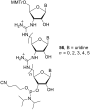
Figure 24: Phosphoramidite building block 56.

Figure 25: Structures of DNGs 57–65.
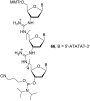
Figure 26: Structure of the phosphoramidite building block 66.
Synthesis in the glycosciences
- Thisbe K. Lindhorst
Beilstein J. Org. Chem. 2010, 6, No. 16, doi:10.3762/bjoc.6.16
- Thisbe K. Lindhorst Otto Diels Institute of Organic Chemistry, Christiana Albertina University of Kiel, Otto-Hahn-Platz 3/4, 24098 Kiel, Germany 10.3762/bjoc.6.16 All cells are coated in carbohydrates and glycoconjugates. Today, after decades where sugars were regarded mainly as a means of energy
Synthesis of a new class of aminocyclitol analogues with the conduramine D-2 configuration
- Latif Kelebekli,
- Yunus Kara and
- Murat Celik
Beilstein J. Org. Chem. 2010, 6, No. 15, doi:10.3762/bjoc.6.15
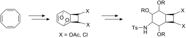
- ]. Carba analogues of oligosaccharides (carbasugars), generated by replacing the endocyclic O-atom in a monosaccharide [1][2][3][4][5][6][7][8][9][10][11], are thought to be better drug candidates than natural sugars, since they are hydrolytically stable. Spurred on by the heightened interest in the design

Graphical Abstract

Scheme 1: Synthesis of bis-carbamate 12 and oxazolidinone 13.

Scheme 2: Mechanism of the palladium-catalyzed ionization/cyclization reaction.

Scheme 3: Synthesis of aminocyclitol analogue 6.

Figure 1: The thermal ellipsoid plot of the single crystal X-ray crystallographic structure of 18.

Scheme 4: Synthesis of oxazolidone 23.

Scheme 5: Mechanism of the palladium-catalyzed ionization/cyclization reaction in dichloro biscarbamate 22.

Scheme 6: Synthesis of dichloroaminocyclitol 7.

Figure 2: 1H NMR NOE spectrum of compound 7.
Recognition properties of receptors consisting of imidazole and indole recognition units towards carbohydrates
- Monika Mazik and
- André Hartmann
Beilstein J. Org. Chem. 2010, 6, No. 9, doi:10.3762/bjoc.6.9
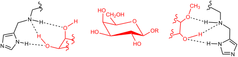
- ][43][48][49][50]). Extractions of sugars 6b, 7b, 8b, 9 and 10 from the solid state into a CDCl3 solution of receptor 4 or 5 (1 mM) provided evidence for strong complexation of β-glucoside 6b and β-galactoside 8b. The extraction of solid methyl α-glucoside 7b, α-galactoside 9 and α-mannoside 10 into a
- CDCl3 solution of receptor 4 or 5 indicated a weaker binding of these sugars than that of 6b and 8b (see Table 1). The extraction experiments indicated that the imidazole-based receptor 4 is a more powerful carbohydrate receptor than the indole-based compound 5. Receptor 4 was able to dissolve about 1
- was carried out on silica gel 60 F254 plates employing chloroform/methanol mixtures as the mobile phase. Melting points are uncorrected. Sugars 6–11, 4(5)-imidazole-carbaldehyde (18) and 3-indole-carbaldehyde (19) are commercially available. General procedure for the synthesis of compounds 4 and 5: To

Graphical Abstract

Figure 1: Examples of hydrogen bonds in the complex of a) galactose-binding protein with D-glucose [3], b) Amara...

Figure 2: Structures of receptors 1–5.

Figure 3: Structures of sugars investigated in this study.
Scheme 1: Reaction conditions: a) AlCl3, CH3CH2Br, 0 °C to r. t., 12 h (85%) [47]; b) 33% HBr in CH3COOH, ZnBr2, ...

Figure 4: Structures of the recently described phenanthroline/aminopyridine-based receptors showing α- vs. β-...

Figure 5: Partial 1H NMR spectra (400 MHz; CDCl3) of receptor 4 (a), 5 (b), 1 (c), and 3 (d) before (bottom) ...

Figure 6: Partial 1H NMR spectra (400 MHz, CDCl3) of 5 after addition of (from bottom to top) 0.00–1.63 equiv...

Figure 7: Energy-minimized structure of the 1:1 a) and 2:1 complex b) formed between receptor 4 and β-galacto...

Figure 8: Examples of hydrogen bonding motifs indicated by molecular modeling studies in the 1:1 complex betw...
Acid- mediated reactions under microfluidic conditions: A new strategy for practical synthesis of biofunctional natural products
- Katsunori Tanaka and
- Koichi Fukase
Beilstein J. Org. Chem. 2009, 5, No. 40, doi:10.3762/bjoc.5.40
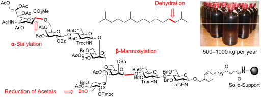
- preparing bioactive natural products [27][28][29][30][31][32][33]. Our successful examples are cation-mediated reactions, such as α-sialylation [28][32], β-mannosylation [31], and reductive opening of the benzylidene acetal groups in sugars [30], for which improved procedure under the microfluidic

Graphical Abstract

Figure 1: Synthetic strategy for asparagine-linked oligosaccharide on solid support and application of microf...

Figure 2: β-Mannosylation using an integrated microfluidic/batch system. Yield and β/α-ratio are analyzed by 1...

Scheme 1: Synthesis of pristane.

Figure 3: Process synthesis of pristane via microfluidic dehydration as a key step.

Scheme 2: Microfluidic dehydration.
Recent progress on the total synthesis of acetogenins from Annonaceae
- Nianguang Li,
- Zhihao Shi,
- Yuping Tang,
- Jianwei Chen and
- Xiang Li
Beilstein J. Org. Chem. 2008, 4, No. 48, doi:10.3762/bjoc.4.48
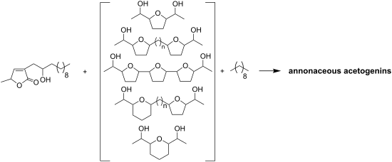
- materials (e.g. amino acids, sugars, tartaric acid, etc.) or on asymmetric reactions {e.g. Sharpless asymmetric epoxidation (AE), Sharpless asymmetric dihydroxylation (AD), diastereoselective Williamson etherification, etc.}. Semi-synthesis of natural ACGs as well as derivatised ACGs (e.g. amines, esters

Graphical Abstract

Scheme 1: Total synthesis of longifolicin by Marshall’s group.

Scheme 2: Total synthesis of corossoline by Tanaka’s group.

Scheme 3: Total synthesis of corossoline by Wu’s group.

Scheme 4: Total synthesis of pseudo-annonacin A by Hanessian’s group.

Scheme 5: Total synthesis of tonkinecin by Wu’s group.

Scheme 6: Total synthesis of gigantetrocin A by Shi’s group.

Scheme 7: Total synthesis of annonacin by Wu’s group.

Scheme 8: Total synthesis of solamin by Kitahara’s group.

Scheme 9: Total synthesis of solamin by Mioskowski’s group.

Scheme 10: Total synthesis of cis-solamin by Makabe’s group.

Scheme 11: Total synthesis of cis-solamin by Brown’s group.

Scheme 12: The formal synthesis of (+)-cis-solamin by Donohoe’s group.

Scheme 13: Total synthesis of cis-solamin by Stark’s group.

Scheme 14: Total synthesis of mosin B by Tanaka’s group.

Scheme 15: Total synthesis of longicin by Hanessian’s group.

Scheme 16: Total synthesis of murisolin and 16,19-cis-murisolin by Tanaka’s group.

Scheme 17: Synthesis of a stereoisomer library of (+)-murisolin by Curran’s group.

Scheme 18: Total synthesis of murisolin by Makabe’s group.

Scheme 19: Total synthesis of reticulatain-1 by Makabe’s group.

Scheme 20: Total synthesis of muricatetrocin C by Ley’s group.

Scheme 21: Total synthesis of (4R,12S,15S,16S,19R,20R,34S)-muricatetrocin (146) and (4R,12R,15S,16S,19R,20R,34S...

Scheme 22: Total synthesis of parviflorin by Hoye’s group.

Scheme 23: Total synthesis of parviflorin by Trost’s group.

Scheme 24: Total synthesis of trilobacin by Sinha’s group.

Scheme 25: Total synthesis of 15-epi-annonin I 181b by Scharf’s group.

Scheme 26: Total synthesis of squamocin A and squamocin D by Scharf’s group.

Scheme 27: Total synthesis of asiminocin by Marshall’s group.

Scheme 28: Total synthesis of asiminecin by Marshall’s group.

Scheme 29: Total synthesis of (+)-(30S)-bullanin by Marshall’s group.

Scheme 30: Total synthesis of uvaricin by the group of Sinha and Keinan.

Scheme 31: Formal synthesis of uvaricin by Burke’s group.

Scheme 32: Total synthesis of trilobin by Marshall’s group.

Scheme 33: Total synthesis of trilobin by the group of Sinha and Keinan.

Scheme 34: Total synthesis of asimilobin by the group of Wang and Shi.

Scheme 35: Total synthesis of squamotacin by the group of Sinha and Keinan.

Scheme 36: Total synthesis of asimicin by Marshall’s group.

Scheme 37: Total synthesis of asimicin by the group of Sinha and Keinan.

Scheme 38: Total synthesis of asimicin by Roush’s group.

Scheme 39: Total synthesis of asimicin by Marshall’s group.

Scheme 40: Total synthesis of 10-hydroxyasimicin by Ley’s group.

Scheme 41: Total synthesis of asimin by Marshall’s group.

Scheme 42: Total synthesis of bullatacin by the group of Sinha and Keinan.

Scheme 43: Total synthesis of bullatacin by Roush’s group.

Scheme 44: Total synthesis of bullatacin by Pagenkopf’s group.

Scheme 45: Total synthesis of rollidecins C and D by the group of Sinha and Keinan.

Scheme 46: Total synthesis of 30(S)-hydroxybullatacin by Marshall’s group.

Scheme 47: Total synthesis of uvarigrandin A and 5(R)-uvarigrandin A by Marshall’s group.

Scheme 48: Total synthesis of membranacin by Brown’s group.

Scheme 49: Total synthesis of membranacin by Lee’s group.

Scheme 50: Total synthesis of rolliniastatin 1 and rollimembrin by Lee’s group.

Scheme 51: Total synthesis of longimicin D by the group of Maezaki and Tanaka.

Scheme 52: Total synthesis of the structure proposed for mucoxin by Borhan’s group.

Scheme 53: Modular synthesis of adjacent bis-THF annonaceous acetogenins by Marshall’s group.

Scheme 54: Total synthesis of 4-deoxygigantecin by Tanaka’s group.

Scheme 55: Total synthesis of squamostatins D by Marshall’s group.

Scheme 56: Total synthesis of gigantecin by Crimmins’s group.

Scheme 57: Total synthesis of gigantecin by Hoye’s group.

Scheme 58: Total synthesis of cis-sylvaticin by Donohoe’s group.

Scheme 59: Total synthesis of 17(S),18(S)-goniocin by Sinha’s group.

Scheme 60: Total synthesis of goniocin and cyclogoniodenin T by the group of Sinha and Keinan.

Scheme 61: Total synthesis of jimenezin by Takahashi’s group.

Scheme 62: Total synthesis of jimenezin by Lee’s group.

Scheme 63: Total synthesis of jimenezin by Hoffmann’s group.

Scheme 64: Total synthesis of muconin by Jacobsen’s group.

Scheme 65: Total synthesis of (+)-muconin by Kitahara’s group.

Scheme 66: Total synthesis of muconin by Takahashi’s group.

Scheme 67: Total synthesis of muconin by the group of Yoshimitsu and Nagaoka.

Scheme 68: Total synthesis of mucocin by the group of Sinha and Keinan.

Scheme 69: Total synthesis of mucocin by Takahashi’s group.

Scheme 70: Total synthesis of (−)-mucocin by Koert’s group.

Scheme 71: Total synthesis of mucocin by the group of Takahashi and Nakata.

Scheme 72: Total synthesis of mucocin by Evans’s group.

Scheme 73: Total synthesis of mucocin by Mootoo’s group.

Scheme 74: Total synthesis of (−)-mucocin by Crimmins’s group.

Scheme 75: Total synthesis of pyranicin by the group of Takahashi and Nakata.

Scheme 76: Total synthesis of pyranicin by Rein’s group.

Scheme 77: Total synthesis of proposed pyragonicin by the group of Takahashi and Nakata.

Scheme 78: Total synthesis of pyragonicin by Rein’s group.

Scheme 79: Total synthesis of pyragonicin by Takahashi’s group.

Scheme 80: Total synthesis of squamostanal A by Figadère’s group.

Scheme 81: Total synthesis of diepomuricanin by Tanaka’s group.

Scheme 82: Total synthesis of (−)-muricatacin [(R,R)-373a] and its enantiomer (+)-muricatacin [(S,S)-373b] by ...

Scheme 83: Total synthesis of epi-muricatacin (+)-(S,R)-373c and (−)-(R,S)-373d by Scharf’s group.

Scheme 84: Total synthesis of (−)-muricatacin 373a and 5-epi-(−)-muricatacin 373d by Uang’s group.

Scheme 85: Total synthesis of four stereoisomers of muricatacin by Yoon’s group.

Scheme 86: Total synthesis of (+)-muricatacin by Figadère’s group.

Scheme 87: Total synthesis of (+)-epi-muricatacin and (−)-muricatacin by Couladouros’s group.

Scheme 88: Total synthesis of muricatacin by Trost’s group.

Scheme 89: Total synthesis of (−)-(4R,5R)-muricatacin by Heck and Mioskowski’s group.

Scheme 90: Total synthesis of muricatacin (−)-373a by the group of Carda and Marco.

Scheme 91: Total synthesis of (−)- and (+)-muricatacin by Popsavin’s group.

Scheme 92: Total synthesis of (−)-muricatacin by the group of Bernard and Piras.

Scheme 93: Total synthesis of (−)-muricatacin by the group of Yoshimitsu and Nagaoka.

Scheme 94: Total synthesis of (−)-muricatacin by Quinn’s group.

Scheme 95: Total synthesis of montecristin by Brückner’s group.

Scheme 96: Total synthesis of (−)-acaterin by the group of Franck and Figadère.

Scheme 97: Total synthesis of (−)-acaterin by Singh’s group.

Scheme 98: Total synthesis of (−)-acaterin by Kumar’s group.

Scheme 99: Total synthesis of rollicosin by Quinn’s group.

Scheme 100: Total synthesis of Rollicosin by Makabe’s group.

Scheme 101: Total synthesis of squamostolide by Makabe’s group.

Scheme 102: Total synthesis of tonkinelin by Makabe’s group.
Part 1. Reduction of S-alkyl- thionocarbonates and related compounds in the presence of trialkylboranes/air
- Jean Boivin and
- Van Tai Nguyen
Beilstein J. Org. Chem. 2007, 3, No. 45, doi:10.1186/1860-5397-3-45
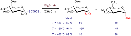
- such as sugars.[3] On the other hand, iodides and S-alkylxanthates are useful compounds that easily produce carbon radicals involved in radical chain reactions (cyclisation, intermolecular addition onto an olefin, etc) in which trapping of the final radical results in the transfer of the iodine atom or
- reduction. In this context, the Surzur-Tanner/Giese rearrangement was particularly illustrative. [22][23][24][25] The 2 → 1 migration of the acyloxy group in glycosyl radicals is well documented and allows an easy preparation of 2-deoxy-sugars. At 20°C in 1,2-dichloroethane, a 50/50 mixture of the "regular

Graphical Abstract

Figure 1: Reaction of α-acylxanthate 1a with 1-decene and Et3B/air.

Figure 2: Xanthates and thionoimidazolides 2–16 and their reduced derivatives.

Scheme 1: Reduction of xanthate 17a at different temperatures with Et3B (5 equiv.)/air.

Scheme 2: Reduction of S-alkylxanthates and O-alkylxanthates.

Scheme 3: Reduction of O-alkyl-S-methyl xanthate 19, thionoimidazolide 21 and iodide 22 by Et3B/air at 20°C.
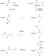
Scheme 4: Products formed through a putative 1,5-hydrogen atom transfer.
An efficient synthesis of tetramic acid derivatives with extended conjugation from L-Ascorbic Acid
- Biswajit K. Singh,
- Surendra S. Bisht and
- Rama P. Tripathi
Beilstein J. Org. Chem. 2006, 2, No. 24, doi:10.1186/1860-5397-2-24

- of multi-step solid and solution phase syntheses exist for the preparation of both achiral and chiral tetramic acid derivatives. [15][16][17][18][19][20][21][22][23][24][25][26][27][28][29][30][31] In an ongoing programme towards the development of new antitubercular agents from sugars, we have been

Graphical Abstract
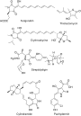
Figure 1: Tetramic acid antibiotics from natural sources.
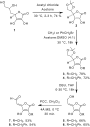
Scheme 1: Synthesis of tetronolactonyl aldehydes from L-ascorbic acid
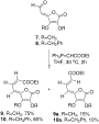
Scheme 2: Synthesis of tetronolactonyl dienyl esters from etronolactonyl aldehydes

Figure 2: H-bonding in tetronolactonyl dienyl esters.

Scheme 3: Synthesis of 5-hydroxy lactams from dienyl tetronic esters

Scheme 4: Synthesis of dienyl tetramic acid from 5- hydroxy lactams
Investigation of acetyl migrations in furanosides
- O. P. Chevallier and
- M. E. Migaud
Beilstein J. Org. Chem. 2006, 2, No. 14, doi:10.1186/1860-5397-2-14
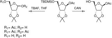
- not occur in compounds 3, 4 or 5 under fluoride-catalysed desilylation conditions and as a result no intramolecular migration products were observed. However, for all sugars, a small proportion of bis-acetylated (compounds c) and non-acetylated (compounds d) compounds have also been isolated (Table 3

Graphical Abstract

Scheme 1: Acetyl migration products upon TBAF/THF treatment

Scheme 2: Synthesis of riboside 1. a) 2,2-Dimethoxypropane, p-toluenesulfonic acid, acetone (65%); b) TBDMSCl...

Scheme 3: Synthesis of xyloside 2 and riboside 3. a) i) acetone, p-toluenesulfonic acid, CuSO4; ii) HCl 0.2 M...

Scheme 4: Synthesis of arabinoside 4. a) HSEt, 6M aq HCl (85%); b) TBDPSCl, imidazole, DMAP, DMF (94%); c) Hg...

Scheme 5: Synthesis of riboside 5. a) BnBr, NaH, THF (82%); b) TBAF, THF (84%); c) PivCl, pyridine/DCM, DMAP ...

Scheme 6: Alkoxide promoted transesterification.
Study of thioglycosylation in ionic liquids
- Jianguo Zhang and
- Arthur Ragauskas
Beilstein J. Org. Chem. 2006, 2, No. 12, doi:10.1186/1860-5397-2-12
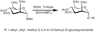
- stability, along with enhanced chemical and thermal stability properties. Recent reports have highlighted the potential of ionic liquids to be used as an ideal solvent for acetylation, ortho-esterification and benzylidenation of sugars,[4][5][6] and for certain glycosylation reactions. Sasaki et al

Graphical Abstract

Figure 1: Structures of common ionic liquids.
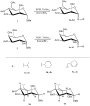
Scheme 1: Glycosylation of 1 and 2 with various glycosyl donors.
Synthesis and glycosidase inhibitory activity of new hexa- substituted C8-glycomimetics
- Olivia Andriuzzi,
- Christine Gravier-Pelletier,
- Gildas Bertho,
- Thierry Prangé and
- Yves Le Merrer
Beilstein J. Org. Chem. 2005, 1, No. 12, doi:10.1186/1860-5397-1-12

- glycosidases have been carried out. For these C8-glycomimetics, weak activities were observed, which can probably be explained by a too high conformational flexibility of such structures. Experimental See Supporting Information File 1. Sugars, iminosugars and carbasugars. Retrosynthetic analysis. 1D proton NMR

Graphical Abstract

Figure 1: Sugars, iminosugars and carbasugars.

Figure 2: Retrosynthetic analysis.

Scheme 1: Reagents and conditions: (a) OsO4, NMO, tBuOH, rt; (b) TFA, H2O, rt.

Scheme 2: Reagents and conditions: (a) mCPBA, CH2Cl2, NaHCO3, rt; (b) see text.

Scheme 3: Reagents and conditions: (a) i : SOCl2, Et3N, CH2Cl2, 0°C; ii : RuCl3, NaIO4, CCl4, CH3CN, 0°C to r...
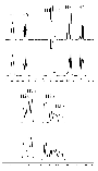
Figure 3: 1D proton NMR spectra of the C8 ring in compound 11 (upper) and the simulated signals (down) on the...

Figure 4: Schematic representation of the NOEs (indicated with arrows) found to deduce the structure of 11. B...

Figure 5: X-ray structure of epoxide 7 (upper) and sulfate 9 (down) solved using SHELXS and anisotropically r...
Towards practical biocatalytic Baeyer- Villiger reactions: applying a thermostable enzyme in the gram- scale synthesis of optically- active lactones in a two-liquid- phase system
- Frank Schulz,
- François Leca,
- Frank Hollmann and
- Manfred T. Reetz
Beilstein J. Org. Chem. 2005, 1, No. 10, doi:10.1186/1860-5397-1-10
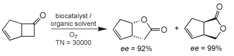
- homogeneity.[56] In order to stabilize the production enzyme in the presence of an organic phase, we evaluated various buffer additives such as sugars, non-ionic detergents, and bovine serum albumin (BSA) which are known to exert beneficial effects on other enzymes.[57] The most pronounced effect on the

Graphical Abstract

Scheme 1:

Figure 1: Evolution of conversion of ketone 1 using PAMO mutant P3 with increasing substrate concentration in...

Scheme 2:

Figure 2: Effect of different solvents on the stability of PAMO, measured as residual activity. For cyclohexa...

Scheme 3:

Figure 3: Conversion during the oxidation of ketone 6 in a two-liquid phase system over the time. The catalys...