

Search results
Search for "validation" in Full Text gives 102 result(s) in Beilstein Journal of Organic Chemistry.
The use of glycoinformatics in glycochemistry
Beilstein J. Org. Chem. 2012, 8, 915–929, doi:10.3762/bjoc.8.104
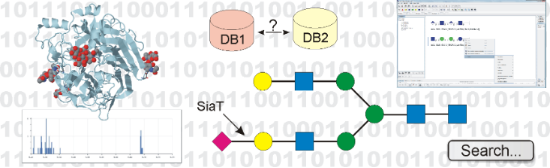
- SugaBase or manually entered from the literature. Glycosciences.DB also contains information on carbohydrate 3D structures that are available in the Protein Data Bank (PDB, [40]). Extraction and validation of carbohydrate data from PDB entries is automated to a large extent and therefore requires only
- significantly lower quality than the protein parts [26][84][85][86]. Reasons for this are both the greater complexity of carbohydrates, and the fact that, while numerous validation tools are available for protein structures [87], only a few programs exist to validate carbohydrate 3D structures. The PDB

An easily accessible sulfated saccharide mimetic inhibits in vitro human tumor cell adhesion and angiogenesis of vascular endothelial cells
Beilstein J. Org. Chem. 2012, 8, 787–803, doi:10.3762/bjoc.8.89
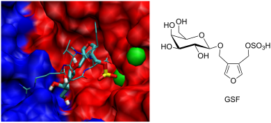
- interaction of GSF with integrins. To find possible binding sites for GSF, we used a “blind docking” approach to screen the protein surface of the extracellular domain of αvβ3, the crystal structure of which was published by Xiong et al. [26]. In an initial validation study, we performed multiple blind


Other Beilstein-Institut Open Science Activities