

Search results
Search for "fluorescence microscopy" in Full Text gives 54 result(s) in Beilstein Journal of Organic Chemistry.
Chemical modification allows phallotoxins and amatoxins to be used as tools in cell biology
Beilstein J. Org. Chem. 2012, 8, 2072–2084, doi:10.3762/bjoc.8.233
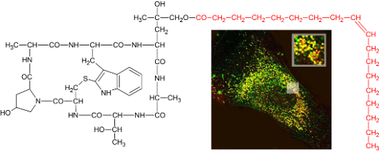
- , albeit under toxic conditions. Using rhodamine-labeled phalloidin, we could also study how membrane-permeable peptides are incorporated into the cell. Immediately after exposure the toxin was located on the plasma membrane of the cells as shown by fluorescence microscopy (Figure 6c), while after 6 h
- -phalloidin (3). (b) Growth inhibition of NIH 3T3 mouse fibroblasts by tetramethylrhodaminyl-phalloidin (3) versus phalloidin (1) after 72 h incubation time. (c) Binding and uptake of tetramethylrhodaminyl-phalloidin (3) in NIH 3T3 mouse fibroblasts after 1, 6 and 24 h, as documented by fluorescence
- microscopy. Chemical structure of amanitin with attachment site for conjugation to internalization-mediating moieties (R1, R3). NIH 3T3 mouse fibroblasts were incubated with various concentrations of α-amanitin and amanitin derivatives. Cell viability was determined after 72 h incubation time by MTT assay




Dimerization of a cell-penetrating peptide leads to enhanced cellular uptake and drug delivery
Beilstein J. Org. Chem. 2012, 8, 1788–1797, doi:10.3762/bjoc.8.204
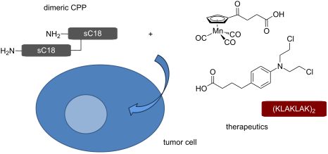
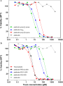
- a dramatically improved capacity to internalize into various cell lines, even primary cells, using flow cytometry and fluorescence microscopy. Cell viability assays indicated increased cytotoxicity of the dimer presumably caused by membrane leakage; however, this effect turned out to be dependent on
- investigated the intracellular distribution pattern of the fluorescently-labeled peptide in HEK-293 by fluorescence microscopy (Figure 2). The punctate uptake pattern speaks in favor of an endocytic internalization mode and is also observed for sC18, which is in line with previous reports [8]. Therefore, the


ROMP-Derived cyclooctene-based monolithic polymeric materials reinforced with inorganic nanoparticles for applications in tissue engineering
Beilstein J. Org. Chem. 2010, 6, 1199–1205, doi:10.3762/bjoc.6.137
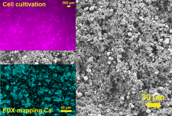
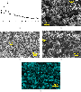
- -derived stromal cells. Each data point is the average of 12 data points. (b) Fluorescence microscopy images of living cells after one and four days of cultivation; cells (human adipose tissue-derived stem cells) grown on a COE-based scaffold reinforced with 12 wt % CaCO3. ROMP-based synthesis of cis-5



Bioorthogonal metabolic glycoengineering of human larynx carcinoma (HEp-2) cells targeting sialic acid
Beilstein J. Org. Chem. 2010, 6, No. 24, doi:10.3762/bjoc.6.24
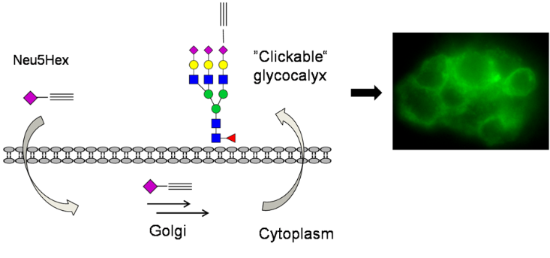
- detectable. In order to analyze the natural background fluorescence of HEp-2, one sample was incubated without any additional carbohydrates. The cells were analyzed by fluorescence microscopy (580 nm for TAMRA staining and at 525 nm for fluorescein). At either wavelength, the negative control does not show
- 2 mM CuSO4, 10 mM sodium ascorbate and 2 mM Tris-[(1-benzyl-1H-1,2,3-triazol-4-yl) methyl]amine (TBTA) in DMSO. After 1 h each well was washed several times with DMSO/water (1:1) and subsequently examined by fluorescence microscopy. Top left: HEp-2 cells incorporated with Ac4GlcNAz 16, labelled with




Other Beilstein-Institut Open Science Activities